Two teeth, seven years: a root canal without a crown, and a filling that saved the nerve
An upper premolar needed a root canal and was protected with a direct composite overlay instead of a crown. A lower molar with deep decay was treated with partial caries removal to keep its nerve alive. Both are healthy seven years later.
From fifth grade to a fellow dentist
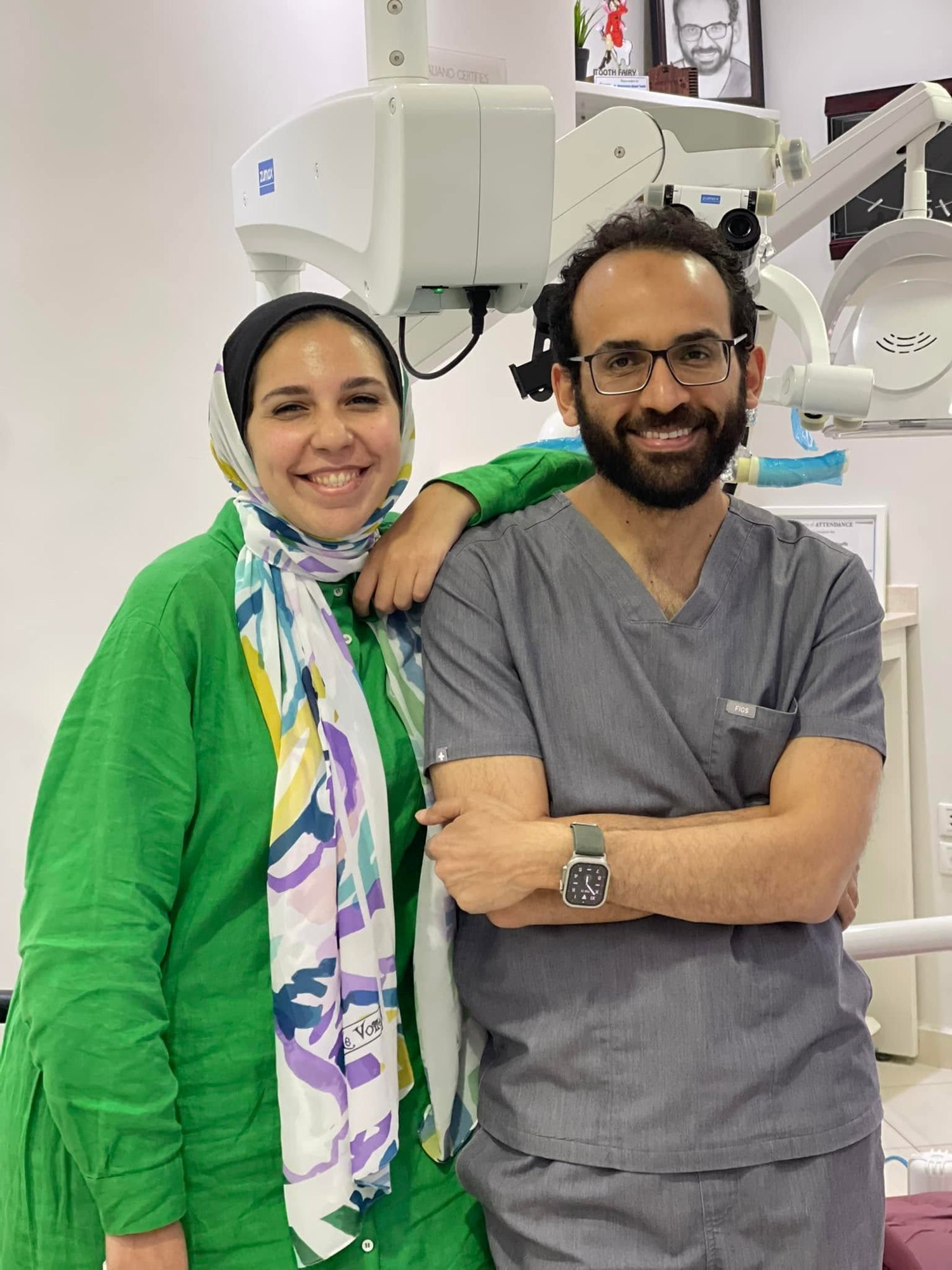
My sister, Dr. Nourhan, started her dental care with me when she was in fifth grade. Today she is a dentist herself. Over the years I have treated almost everything for her: root canals, gum treatment, implants, composite restorations and crowns.
Seven years ago, two of her teeth needed treatment at the same time, and each had a different story.
The upper premolar: a root canal without a crown
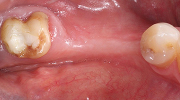
The decay in her upper left first premolar was deep, below the gum line, and had reached the nerve. The usual answer in a case like this is to cut the tooth down for a crown.
We chose a path that keeps the natural tooth: we rebuilt the missing part before the root canal, completed the root canal, and then covered the tooth with a direct composite restoration that protects its cusps, without cutting away any healthy tooth.
The lower molar: a filling that kept the nerve alive
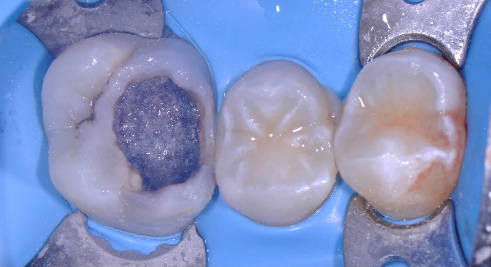
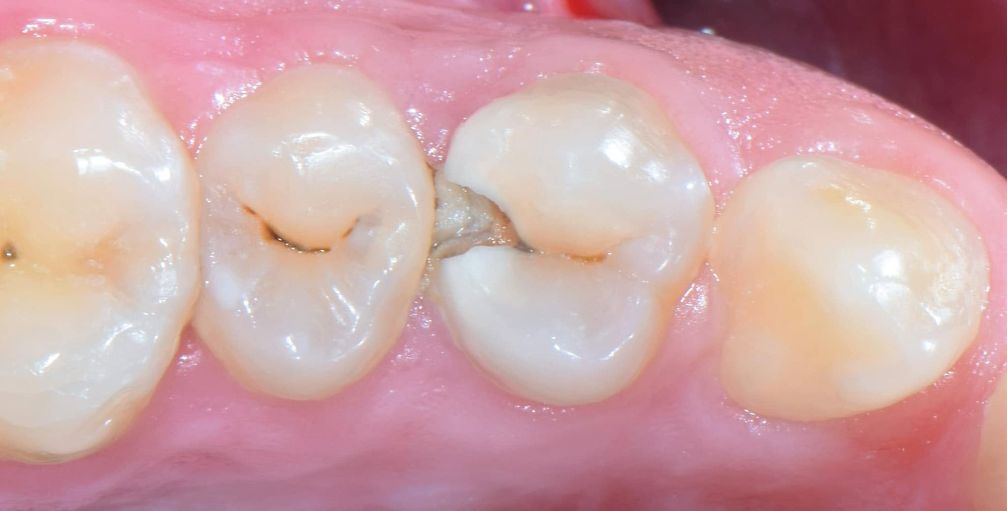
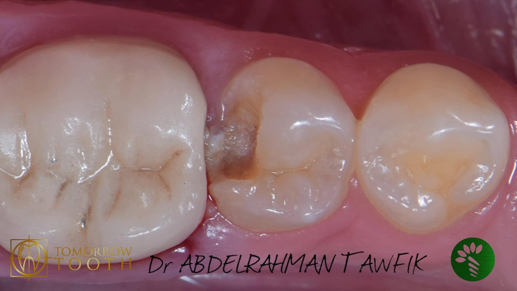
The lower molar had deep decay very close to the nerve. Removing every last bit of it would have exposed the nerve and led to a root canal.
So we removed the decay selectively: the edges were cleaned completely, a thin layer was left over the nerve, and the tooth was sealed with a tight restoration. The nerve stayed alive, and a root canal was avoided altogether.
Seven years later
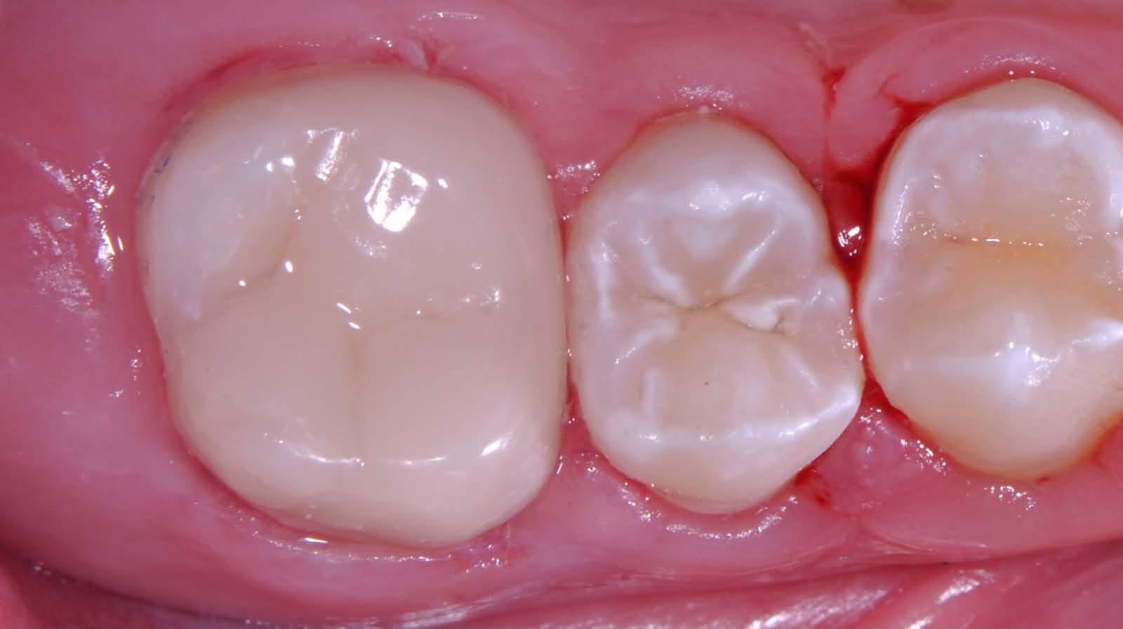
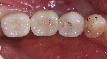
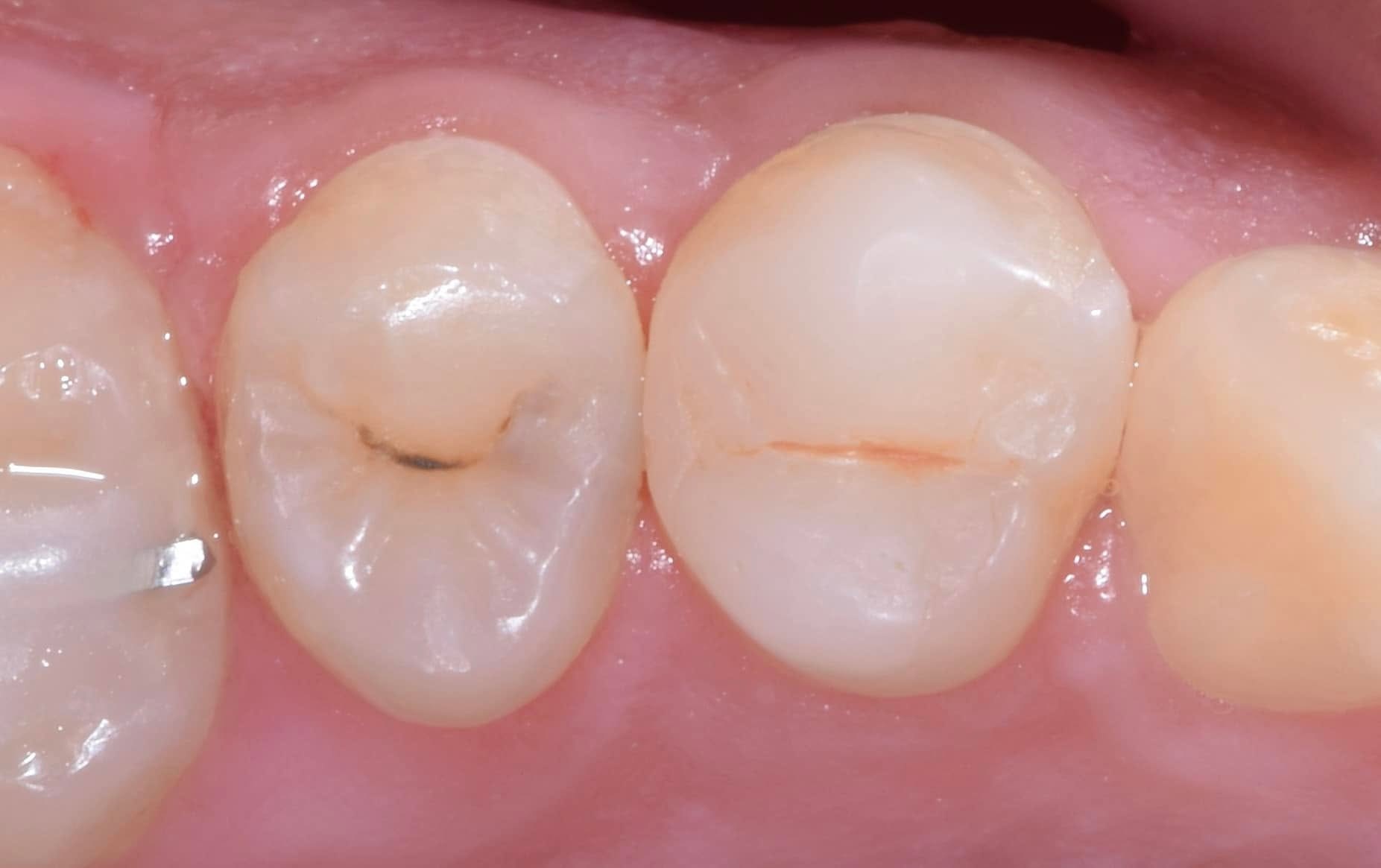
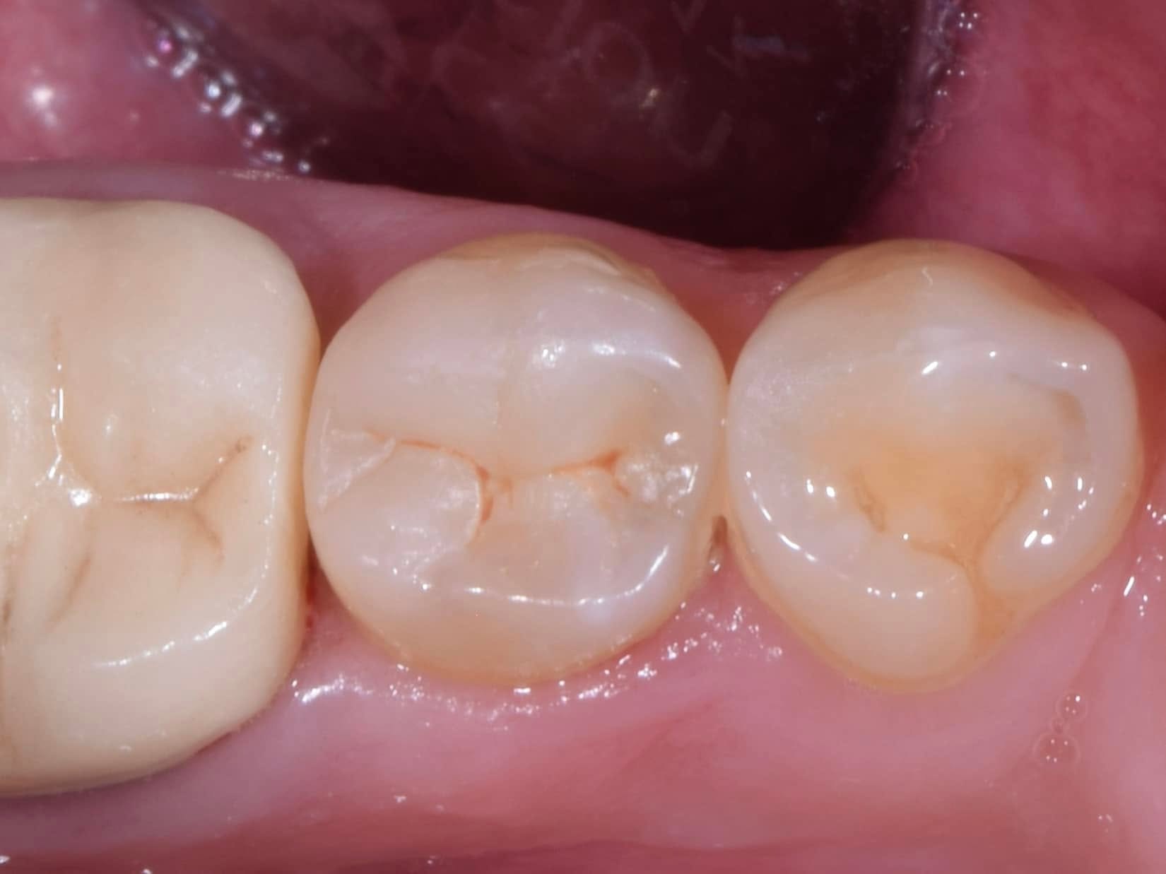
Today both restorations are in place, both teeth are healthy, the gums are healthy, and the lower molar still has its natural, living nerve.
Common questions
- Do you always need a crown after a root canal? Not always. If enough of the tooth remains, it can be protected with a restoration that covers its cusps.
- Can a root canal be avoided when decay is deep? Often yes, if the nerve is healthy and there is no lingering pain. Partial caries removal keeps the nerve alive.
- How long do composite fillings last in back teeth? Most last more than ten years with good care and regular check-ups.
- Is it painful? It is done under local anaesthesia, and you feel no pain during treatment.
Clinical details for dentists
1 Upper premolar: pre-op, deep proximal caries below the gum line 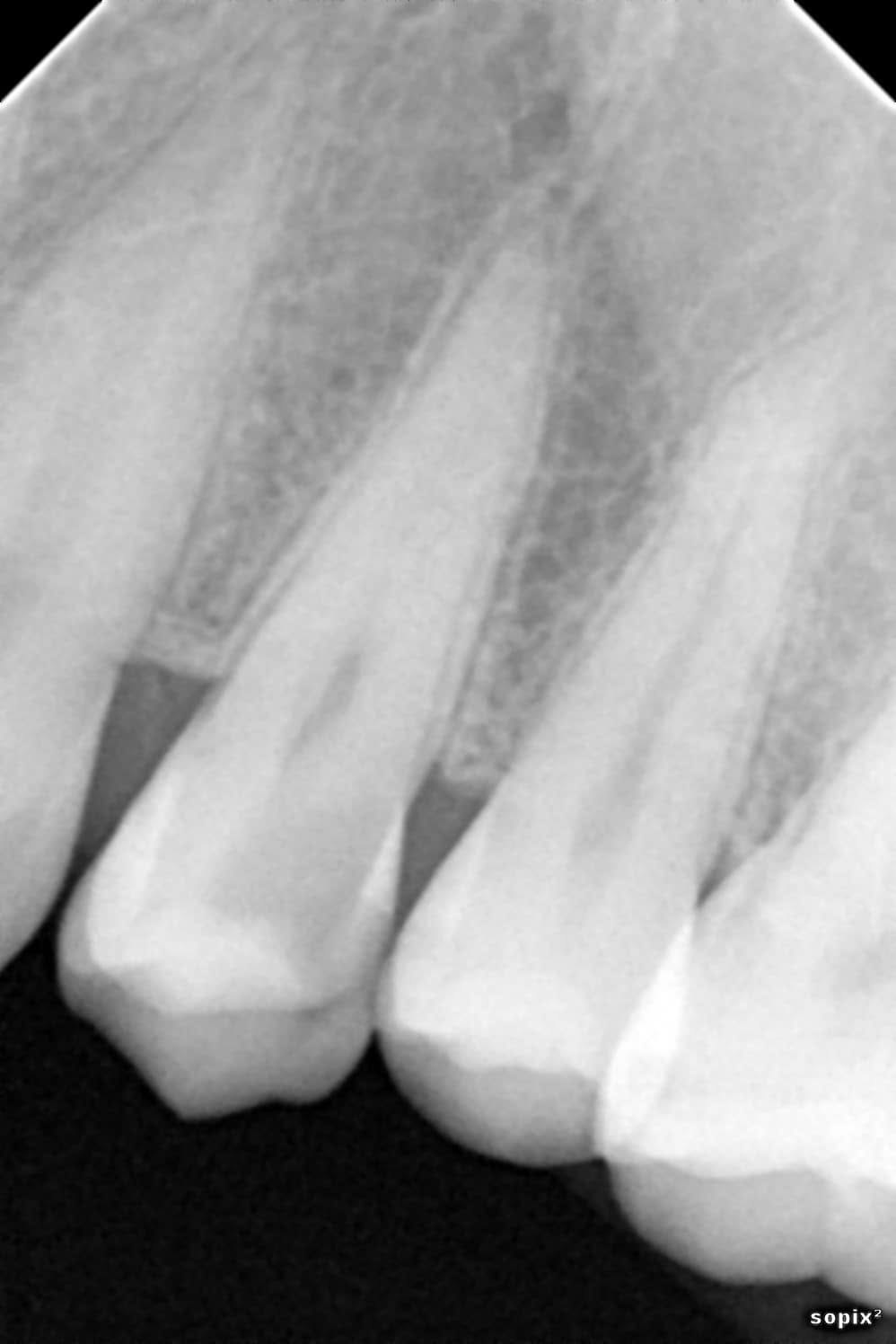
2 Upper premolar: pre-operative radiograph 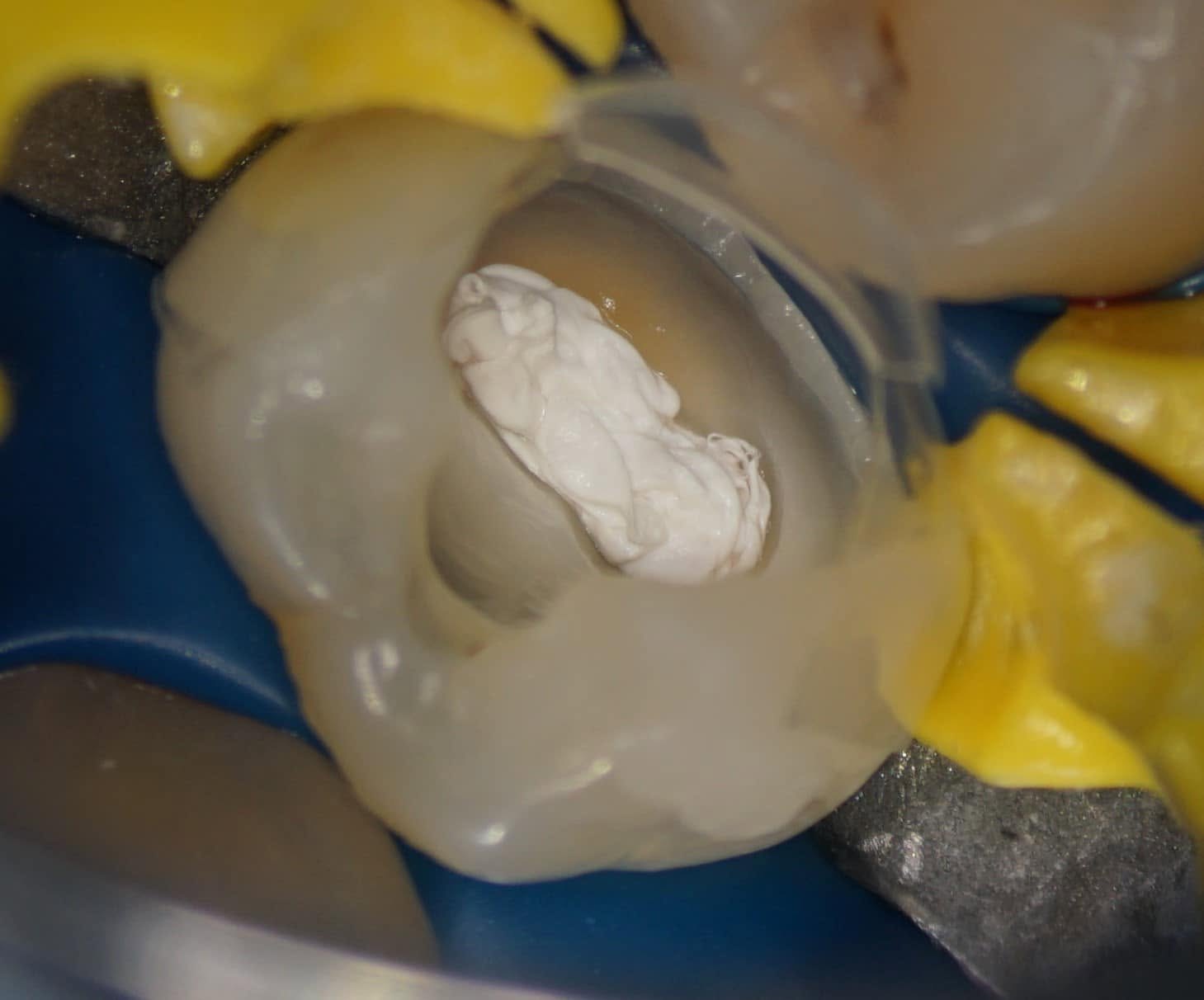
3 Upper premolar: rubber dam and pre-endodontic build-up 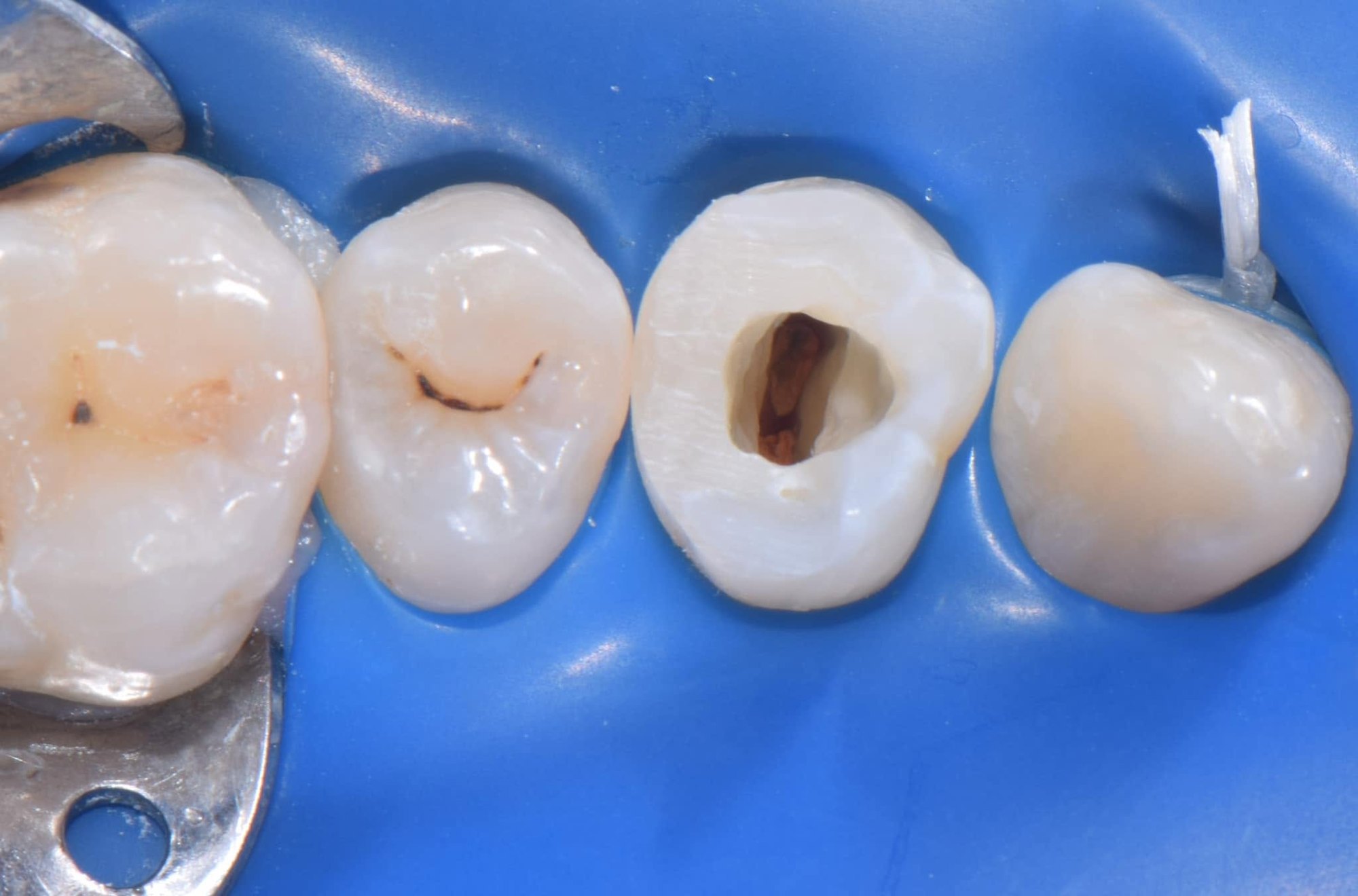
4 Upper premolar: access cavity after the build-up 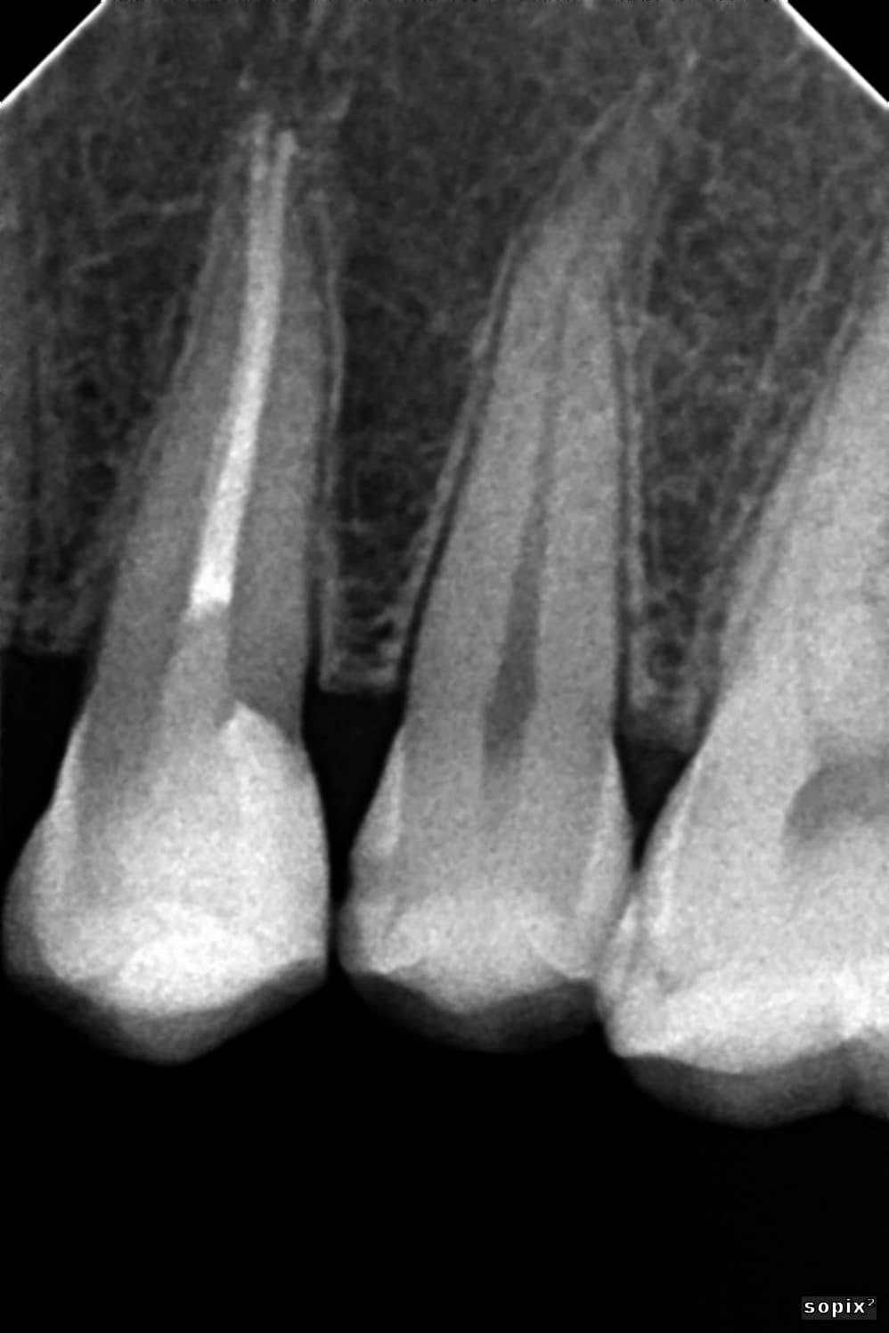
5 Upper premolar: root canal obturation (radiograph) 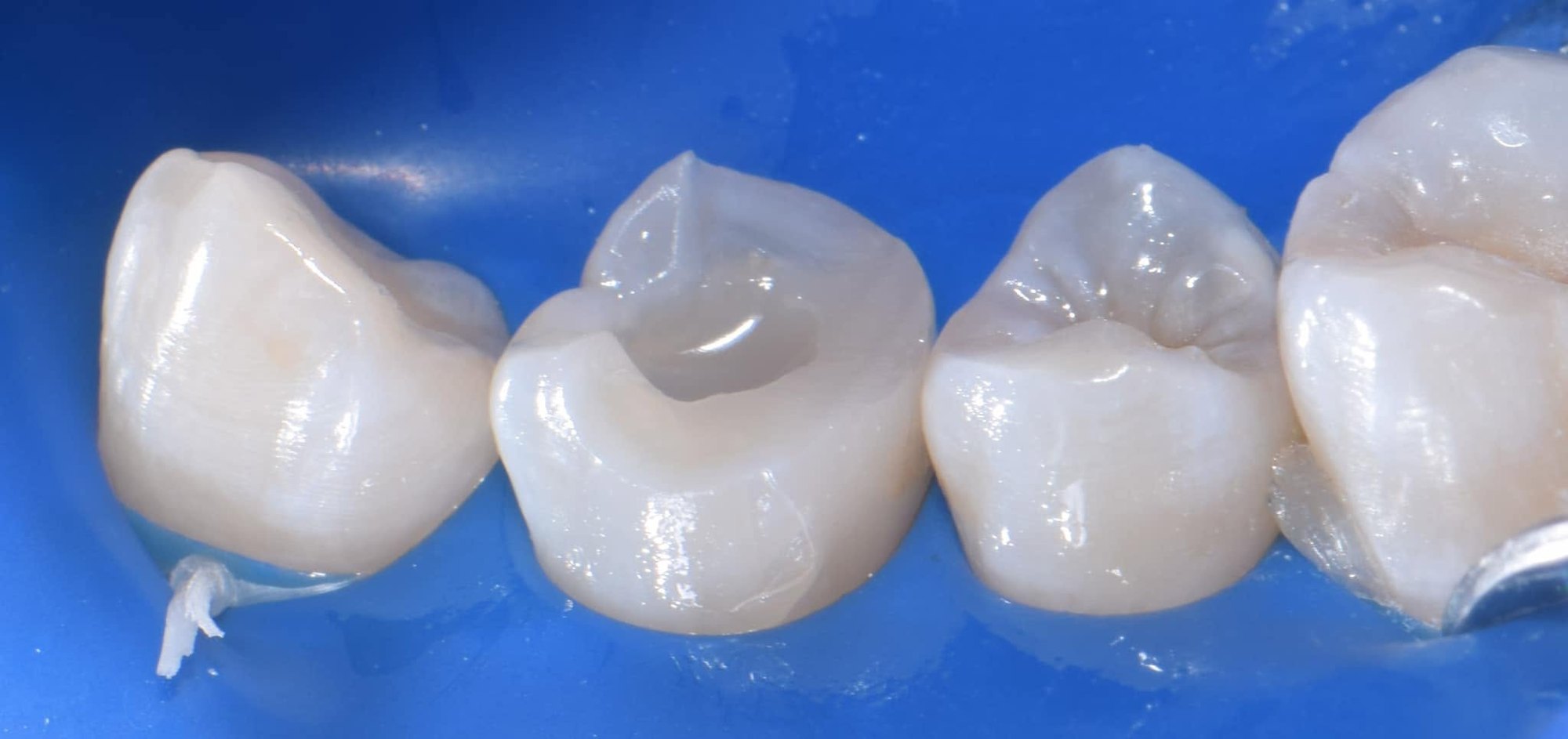
6 Upper premolar: direct cuspal-coverage composite under rubber dam 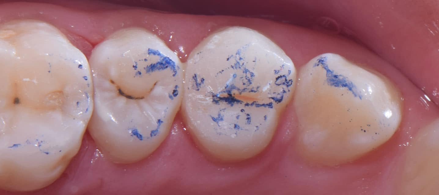
7 Upper premolar: occlusion check 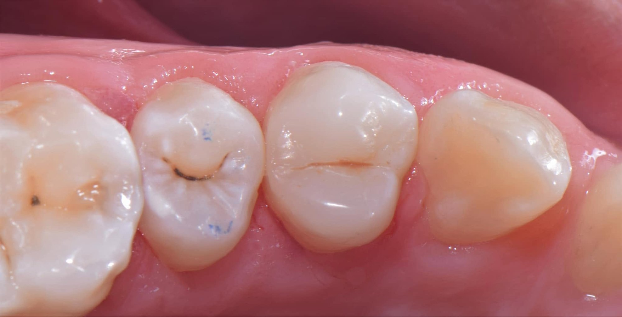
8 Upper premolar: final restoration 9 Upper premolar: 7-year follow-up 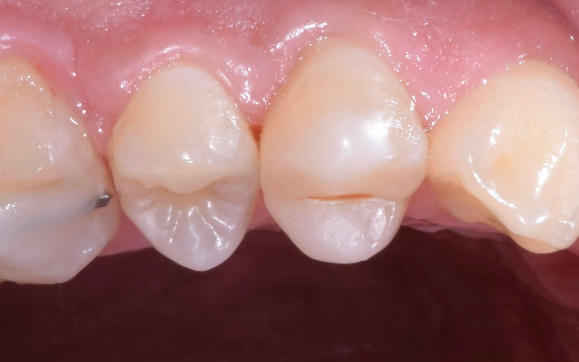
10 Upper premolar: 7-year follow-up 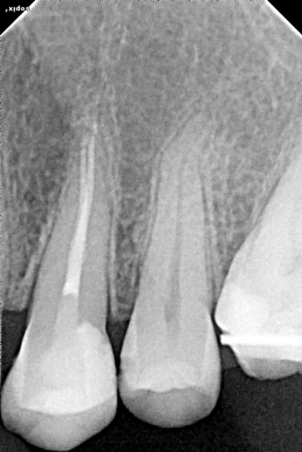
11 Upper premolar: 7-year follow-up radiograph 12 Lower molar: pre-op, deep caries close to the pulp 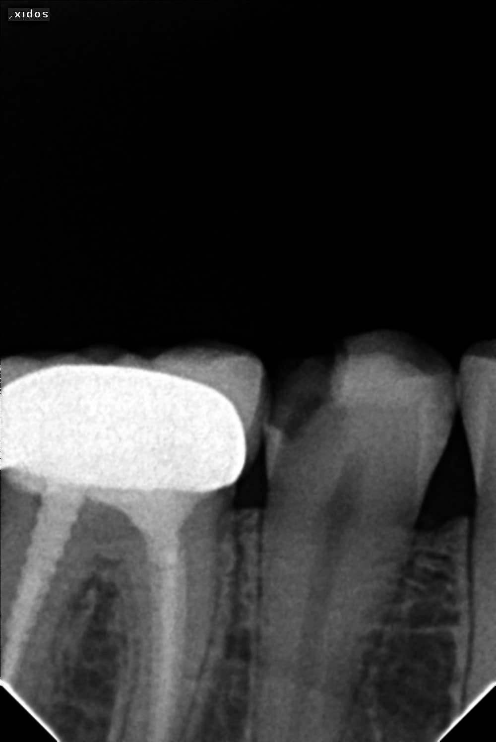
13 Lower molar: pre-operative radiograph 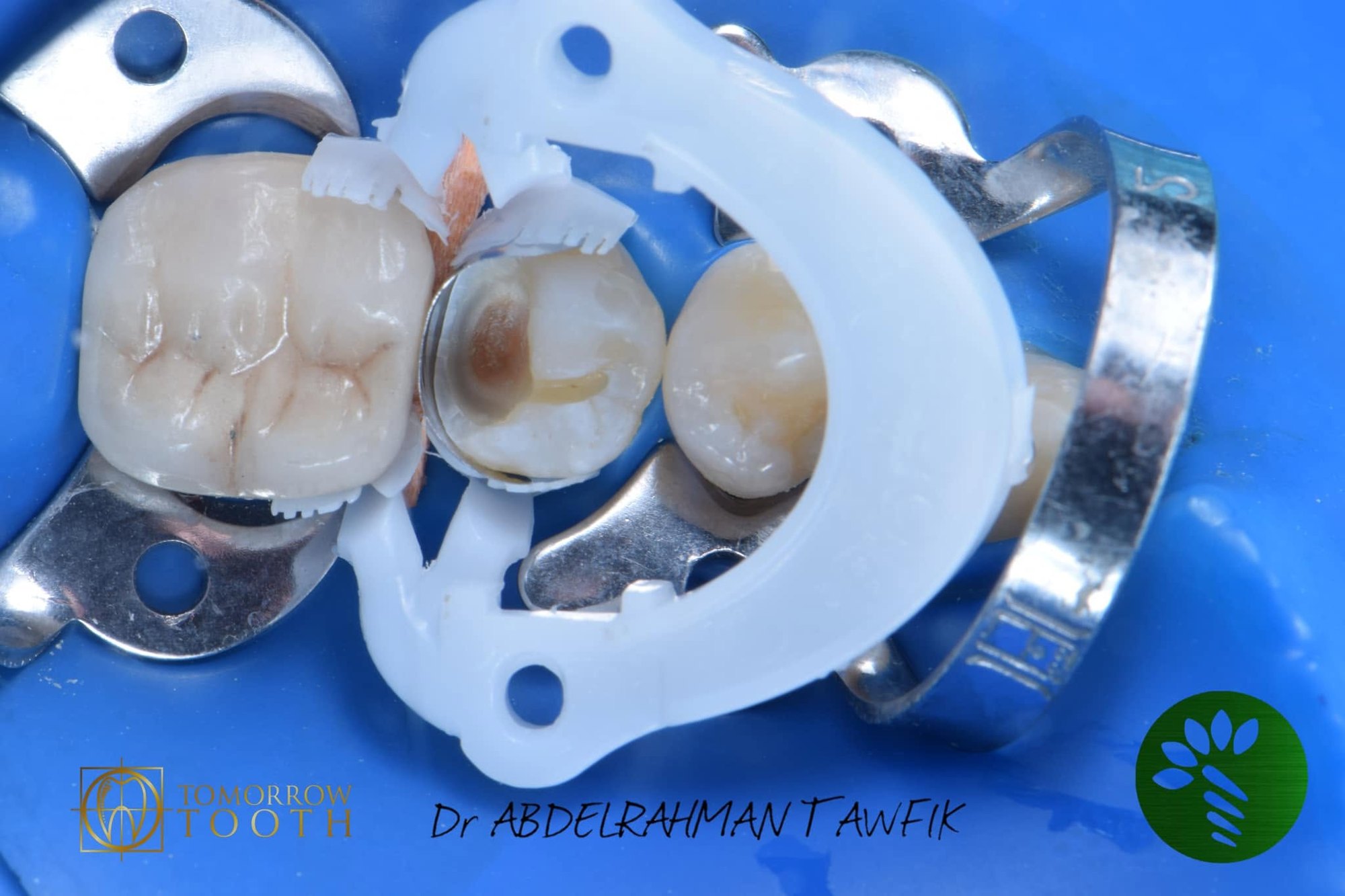
14 Lower molar: multiple-tooth isolation, partial caries removal 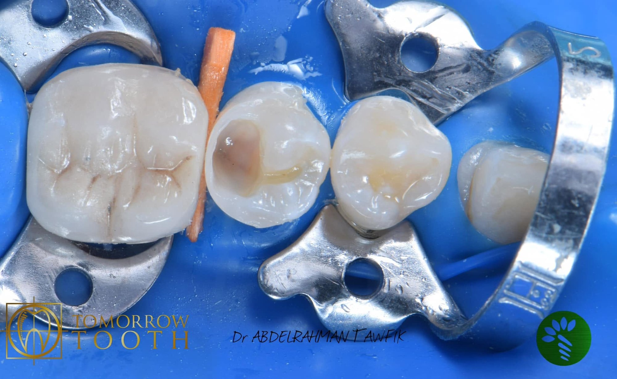
15 Lower molar: proximal wall with TOR VM sectional matrix and Bioclear ring (snowplow) 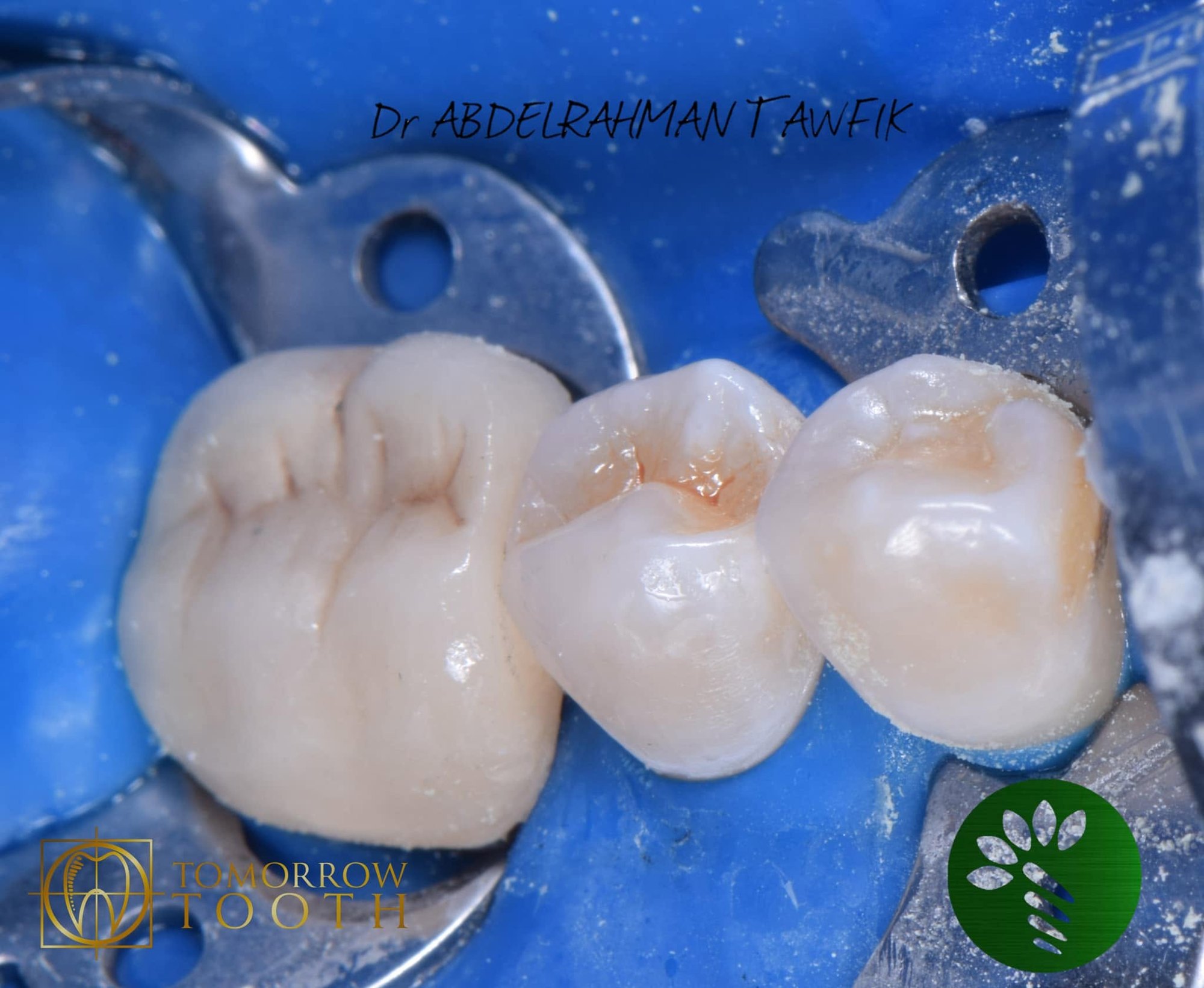
16 Lower molar: occlusal anatomy under rubber dam 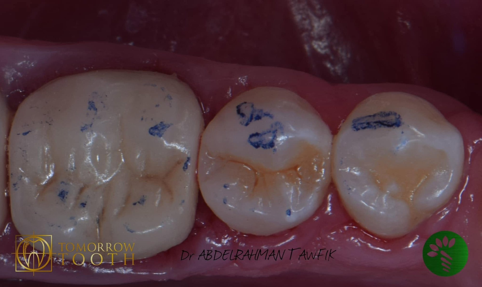
17 Lower molar: occlusion check 18 Lower molar: 7-year follow-up 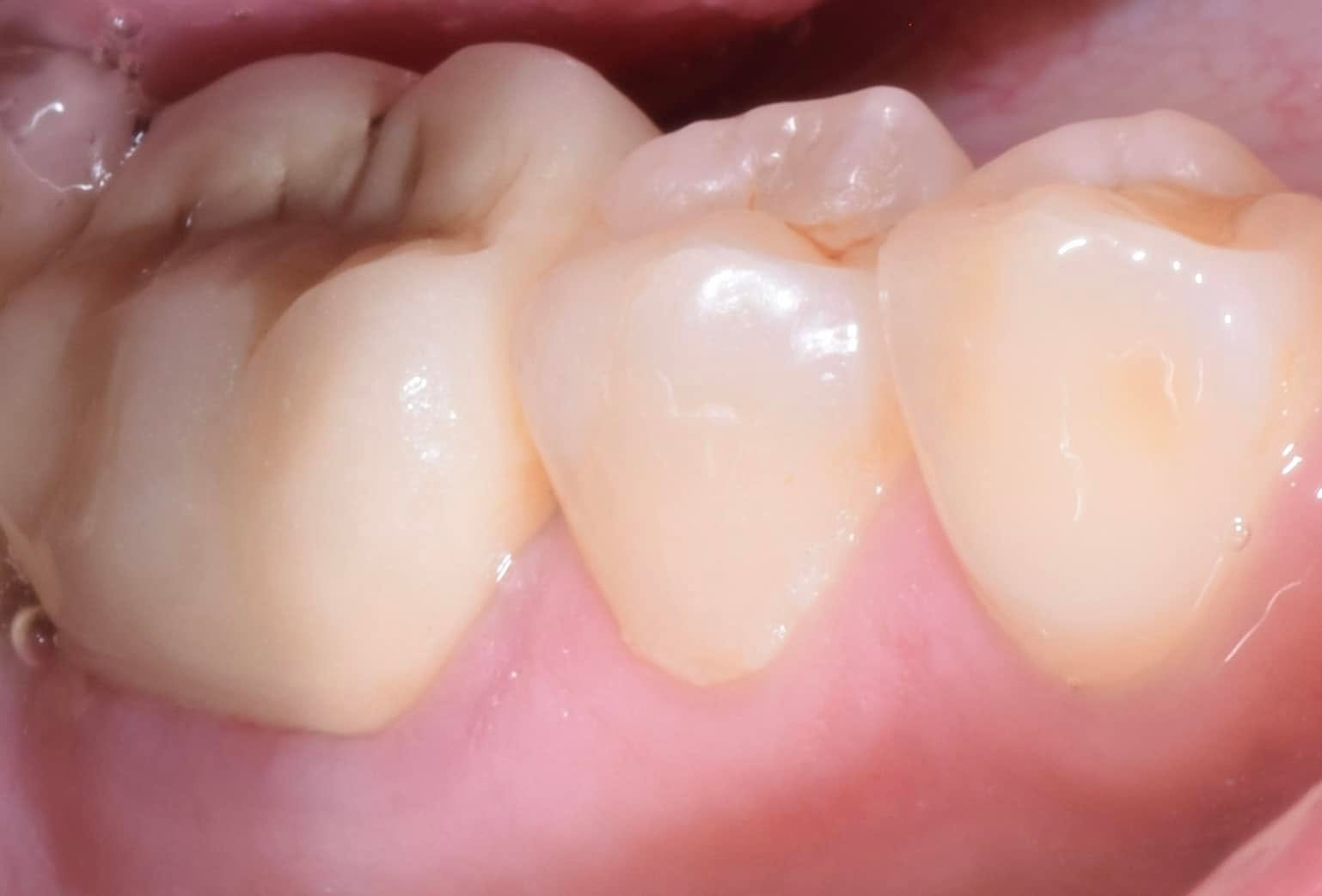
19 Lower molar: 7-year follow-up 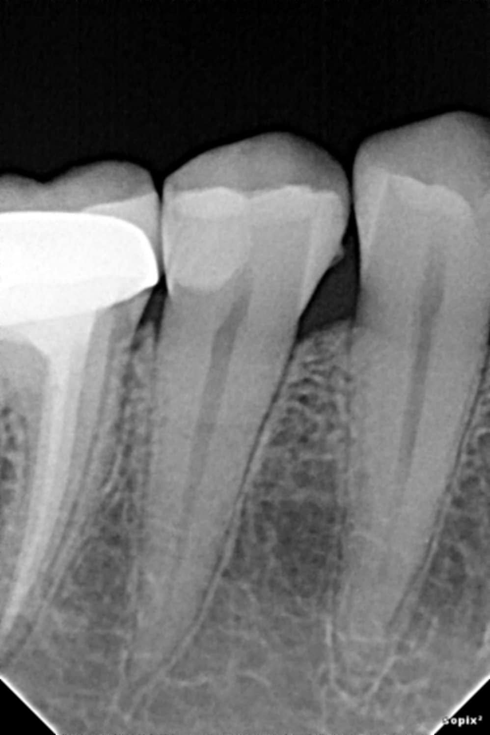
20 Lower molar: 7-year follow-up radiograph
Two teeth, two strategies: 7-year follow-up
- Tooth 1: maxillary left first premolar (#24). Pre-endodontic build-up, root canal treatment and direct cuspal-coverage composite (direct overlay).
- Tooth 2: mandibular molar. Partial (selective) caries removal to preserve pulp vitality, then a direct composite with the proximal wall built first.
Tooth 1: maxillary first premolar
- Deep proximal caries extending subgingivally, with pulp involvement.
- Rubber dam isolation.
- Pre-endodontic build-up with deep margin relocation: Bioclear Biofit matrix, Bioclear Diamond Wedge and Bioclear Twin Ring.
- Snowplow technique at the cervical floor: an uncured layer of flowable composite, with the paste composite pushed into it and both light-cured together.
- Root canal treatment, obturation with a resin-based sealer.
- Definitive direct composite with cuspal coverage (direct overlay).
Tooth 2: mandibular molar
- Deep caries close to the pulp, vital pulp, no signs of irreversible pulpitis.
- Multiple-tooth rubber dam isolation.
- Partial (selective) caries removal: peripheral dentine and enamel margins cleaned to hard tissue, soft dentine left over the pulp to avoid exposure.
- Proximal wall built first with the snowplow technique, using a TOR VM sectional matrix and a Bioclear ring.
- Occlusal anatomy built up, occlusion checked, finished and polished.
Materials
- 3M Filtek Z350 XT composite
- 3M Scotchbond adhesive
- Resin-based endodontic sealer (tooth 1)
Rationale
- Build-up before endo: a predictable rubber-dam seal, a stable reference for working length, and a sealed chamber between visits. Relocating the deep margin follows the deep margin elevation principle, with 95.9% survival reported up to 12 years (Bresser et al., 2019).
- Direct overlay instead of a crown: preserves sound tooth structure. In a randomized trial on endodontically treated premolars, failure rates of direct composite were not significantly different from full crowns (Mannocci et al., 2002). Bruxism is a major risk factor, so case selection matters (Abu-Awwad et al., 2025).
- Coronal seal: the quality of the coronal restoration strongly influences periapical health (Ray & Trope, 1995).
- Partial caries removal: avoiding pulp exposure is the key to keeping the pulp vital. International consensus recommends selective removal in deep lesions (Schwendicke et al., 2016). In a 5-year randomized trial, restorations placed over soft dentine survived as well as those after stepwise excavation (Jardim et al., 2020).
- Longevity: posterior composites show annual failure rates of 1.8% at 5 years and 2.4% at 10 years (Opdam et al., 2014).
Clinical tips
- Rubber dam at every step, including multiple-tooth isolation when restoring adjacent teeth.
- In deep vital teeth, protect the pulp: clean the periphery, not the floor.
- Build the proximal wall first, then the occlusal anatomy.
- Snowplow at the cervical floor for adaptation.
- Use cuspal coverage when the walls are thin.
- Check the occlusion carefully, and recall regularly.
References
- Opdam NJ, et al. Longevity of posterior composite restorations: a systematic review and meta-analysis. J Dent Res. 2014;93(10):943–949.
- Mannocci F, et al. Three-year clinical comparison of survival of endodontically treated teeth restored with either full cast coverage or with direct composite restoration. J Prosthet Dent. 2002.
- Abu-Awwad M, et al. Direct restorations versus full crowns in endodontically treated molar teeth: a three-year randomized clinical trial. J Dent. 2025;156:105699.
- Bresser RA, et al. Up to 12 years clinical evaluation of 197 partial indirect restorations with deep margin elevation in the posterior region. J Dent. 2019;91:103227.
- Ray HA, Trope M. Periapical status of endodontically treated teeth in relation to the technical quality of the root filling and the coronal restoration. Int Endod J. 1995;28(1):12–18.
- Schwendicke F, et al. Managing carious lesions: consensus recommendations on carious tissue removal. Adv Dent Res. 2016;28(2):58–67.
- Jardim JJ, et al. Restorations after selective caries removal: 5-year randomized trial. J Dent. 2020;99:103416.
Book a Consultation
Leave your details and our team will contact you to arrange your visit — whether you live in Egypt or are travelling in for treatment.